Detecting Nuclei
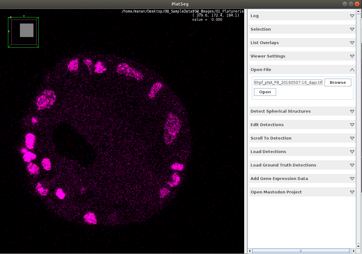
Open Image
Select card Open File
Browse for Image on your local workstation
Open the selected file
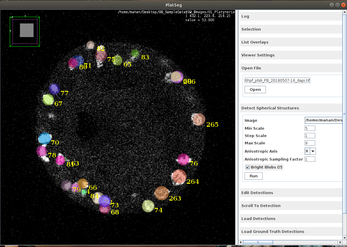
Local Minima in the 4D space
Select card Detect Spherical Structures
Set Min Scale = 5
Set Step Scale = 1
Set Max Scale = 9
Set Anisotropic Axis = X for in-situ embryo and Z for live embryo
Set Anisotropic Sampling Factor = 1 for in-situ embryo and 5 for live embryo
Ensure Bright Blobs checkbox is ticked
Press Run
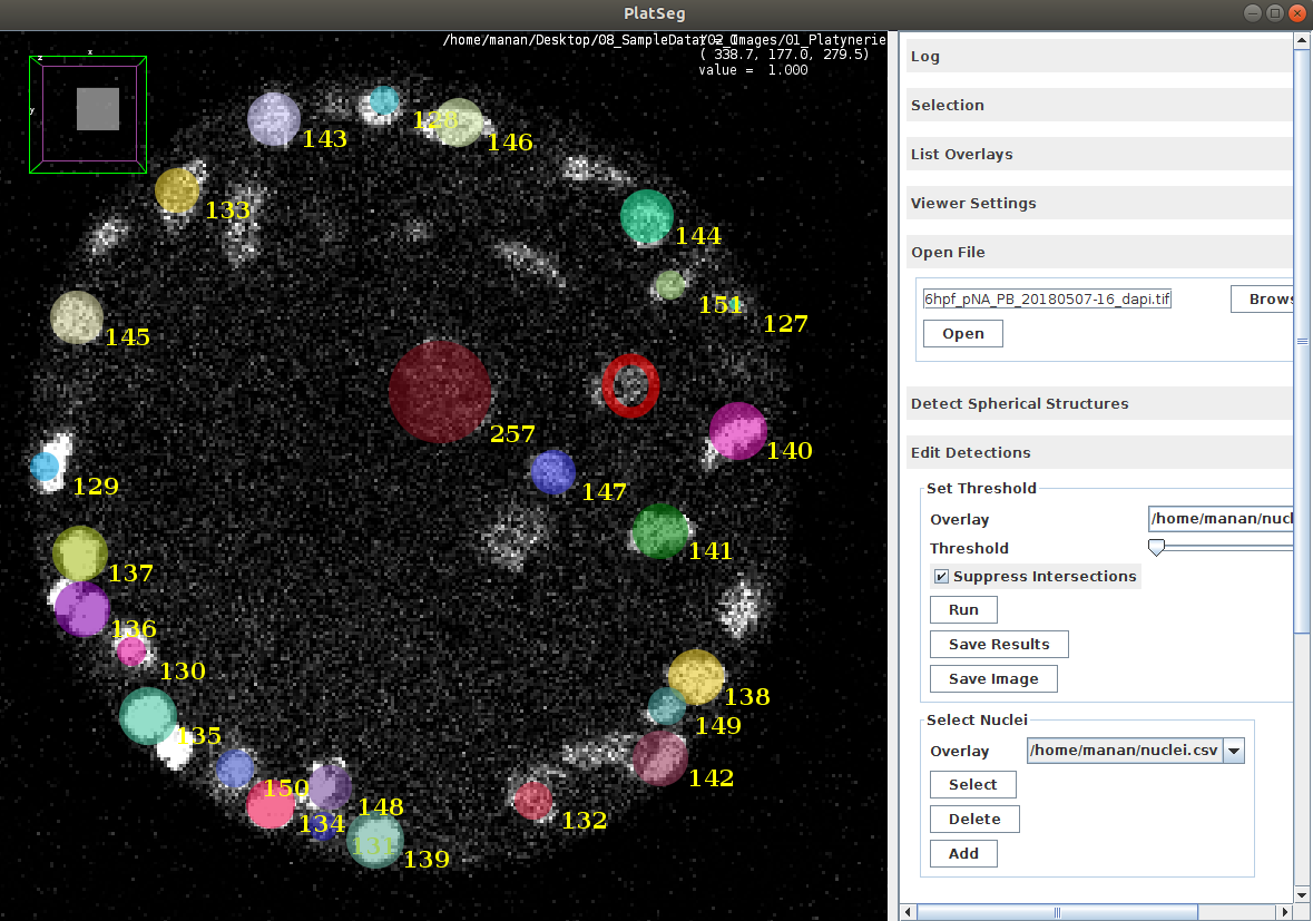
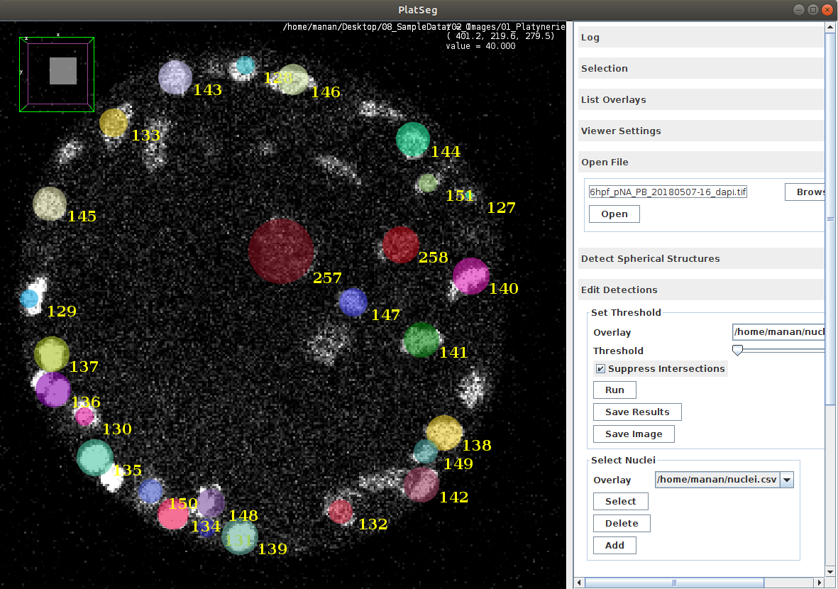
Edit Nuclei
Select card Edit Detections
Press Run
Visually inspect the results
At sites of over-detection, select card Select Nuclei
Click on over-segmented detection and press Delete
At sites of under-detection, select card Select Nuclei
Drag, form an Oval and release. Next click Add
When satisfied, press Save Results
Load Previous Detections
Open Image by using the card Open File
Select card Load Ground Truth Detections
Browse for previously saved csv results file
Click Open to load results file